UCSF-PDGM | The University of California San Francisco Preoperative Diffuse Glioma MRI
DOI: 10.7937/tcia.bdgf-8v37 | Page Accessibility: Public | Collection
| Location | Species | Subjects | Data Types | Cancer Types | Size | Status | Updated | |
|---|---|---|---|---|---|---|---|---|
| Brain | Human | 495 | MR | Diffuse Glioma | Clinical, Genomics, Image Analyses | Public, Complete | 2023/09/13 |
Summary
Summary
Introduction
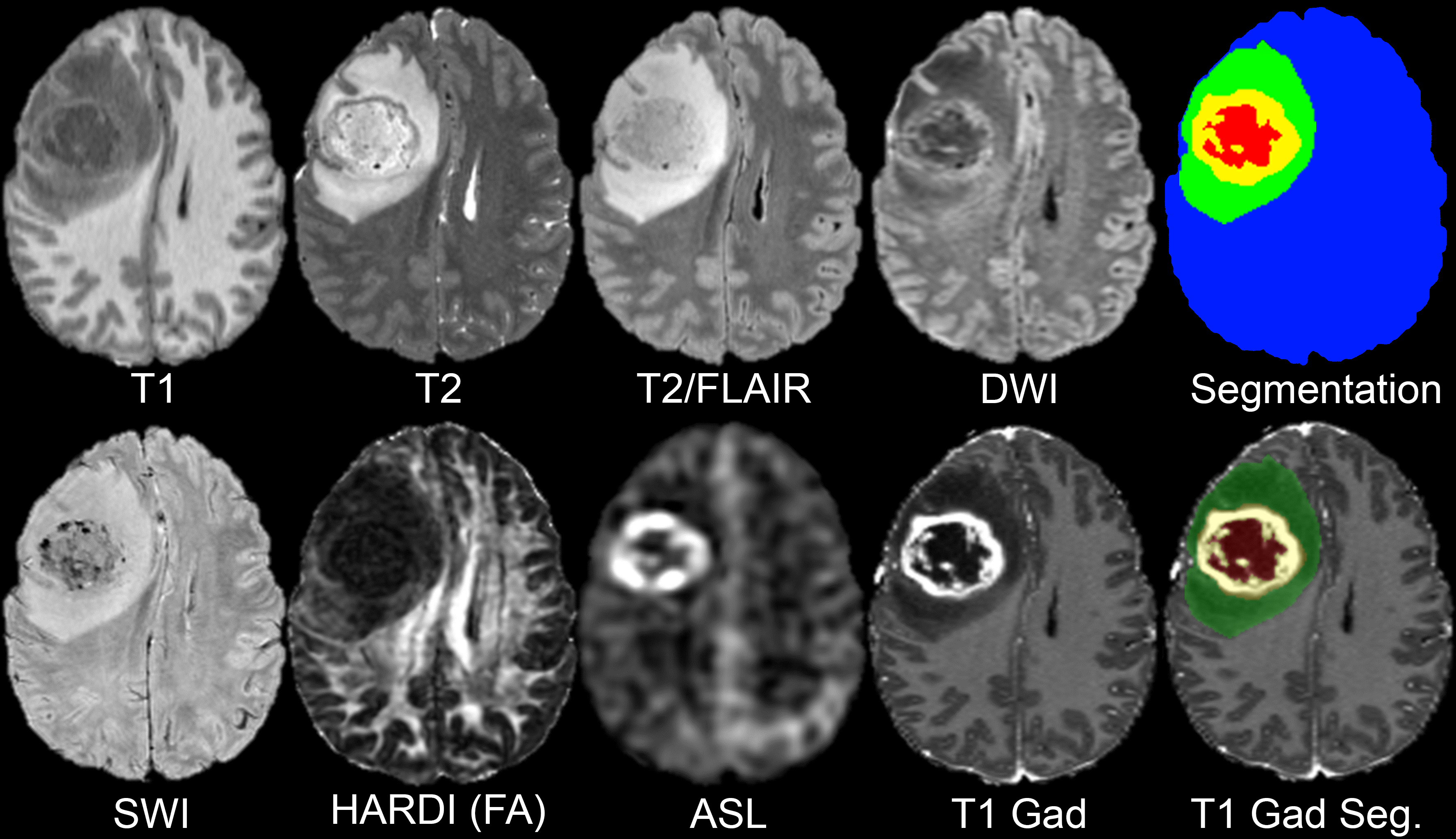
MRI-based artificial intelligence (AI) research on patients with brain gliomas has been rapidly increasing in popularity in recent years in part due to a growing number of publicly available MRI datasets. Notable examples include The Cancer Genome Atlas Glioblastoma dataset (TCGA-GBM) consisting of 262 subjects and the International Brain Tumor Segmentation (BraTS) challenge dataset consisting of 542 subjects (including 243 preoperative cases from TCGA-GBM). The public availability of these glioma MRI datasets has fostered the growth of numerous emerging AI techniques including automated tumor segmentation, radiogenomics, and MRI-based survival prediction. Despite these advances, existing publicly available glioma MRI datasets have been largely limited to only 4 MRI contrasts (T2, T2/FLAIR, and T1 pre- and post-contrast) and imaging protocols vary significantly in terms of magnetic field strength and acquisition parameters. Here we present the University of California San Francisco Preoperative Diffuse Glioma MRI (UCSF-PDGM) dataset. The UCSF-PDGM dataset includes 501 subjects with histopathologically-proven diffuse gliomas who were imaged with a standardized 3 Tesla preoperative brain tumor MRI protocol featuring predominantly 3D imaging, as well as advanced diffusion and perfusion imaging techniques. The dataset also includes isocitrate dehydrogenase (IDH) mutation status for all cases and O[6]-methylguanine-DNA methyltransferase (MGMT) promotor methylation status for World Health Organization (WHO) grade III and IV gliomas. The UCSF-PDGM has been made publicly available in the hopes that researchers around the world will use these data to continue to push the boundaries of AI applications for diffuse gliomas.
Acknowledgements
We would like to acknowledge the individuals and institutions that have provided data for this collection:
Research was supported by the National Institutes of Health Ruth L. Kirschstein Institutional National Research Service Award under award number T32EB001631 and by the RSNA Research & Education Foundation under grant number RR2011. The content is solely the responsibility of the authors and does not necessarily represent the official views of the RSNA R&E Foundation.
Data Access
Click the Versions tab for more info about data releases.
Please contact [email protected] with any questions regarding usage.
| Title | Data Type | Format | Access Points | License | |||
|---|---|---|---|---|---|---|---|
| Images and Annotations | MR | NIFTI | Requires IBM-Aspera-Connect plugin |
501 | 11,523 | CC BY 4.0 | |
| Clinical data | CSV | CC BY 4.0 | |||||
| Clinical metadata glossary | CC BY 4.0 | ||||||
| bval file | CC BY 4.0 | ||||||
| bvec file | CC BY 4.0 |
Detailed Description
*Note: it was discovered that some ID are followup imaging of others, therefore the true number of patients in this Collection is 495 , the pixels have not been changed:
UCSF-PDGM-0433 was imaged 7 days later as UCSF-PDGM-0315 (version 3 files reflect this change by renaming UCSF-PDGM-0315 to UCSF-PDGM-0433_FU007d)
UCSF-PDGM-0431 was imaged 1 days later as UCSF-PDGM-0278 (version 3 files reflect this change by renaming UCSF-PDGM-0278 to UCSF-PDGM-0431_FU001d)
UCSF-PDGM-0396 was imaged 175 days later as UCSF-PDGM-0175 (version 3 files reflect this change by renaming UCSF-PDGM-0175 to UCSF-PDGM-0396_FU175d)
UCSF-PDGM-0429 was imaged 3 days later as UCSF-PDGM-0138 (version 3 files reflect this change by renaming UCSF-PDGM-0138 to UCSF-PDGM-0429_FU003d)
UCSF-PDGM-0409 was imaged 1 days later as UCSF-PDGM-0181 (version 3 files reflect this change by renaming UCSF-PDGM-0181 to UCSF-PDGM-0409_FU001d)
UCSF-PDGM-0391 was imaged 16 days later as UCSF-PDGM-0289 (version 3 files reflect this change by renaming UCSF-PDGM-0289 UCSF-PDGM-0391_FU016d)
All image data have been “skull stripped”, deidentified, pre-processed per the methods section of our abstract, and converted to NIfTI format. We cannot provide original DICOM data, however, these pre-processed files have been prepared to facilitate the type of research that this dataset is intended for. Publicly available deep-learning algorithm for performing this process is here: https://www.github.com/ecalabr/brain_mask/.
Glossary of abbreviations: UCSF-PDGM-metadata_glossary.csv
| Term | Represents | Values |
|---|---|---|
| ID | DICOM (0010,0020) PatientID | |
| Sex | DICOM (0010,0040) Patient Sex | M,F |
| Age at MRI | Age in years at time of MR imaging | |
| WHO CNS Grade | Grade per the 2021 World Health Organization Classification of Tumors of the Central Nervous System (WHO CNS 2021) (https://doi.org/10.1093/neuonc/noab106 ) | 2,3,4 |
| Final pathologic diagnosis (WHO 2021) | Final (integrated) pathologic diagnosis per the 2021 World Health Organization Classification of Tumors of the Central Nervous System (WHO CNS 2021) ( https://doi.org/10.1093/neuonc/noab106 ) |
|
| MGMT status | O6-methylguanine-DNA methyltransferase status – clinical interpretation of the MGMT index described below. | negative, positive, indeterminate |
| MGMT index | O6-methylguanine-DNA methyltransferase methylation index (in house method developed by UCSF clinical labs, https://genomics.ucsf.edu/content/mgmt-promoter-methylation-assay ) where numeric values 0-17 indicate the number of promoter methylation sites. | 0-17, blank |
| 1p/19q | presence of codeletion of 1p and 19q genes, assayed by fluorescent in-situ hybridization | intact, co-deletion, relative co-deletion, unknown |
| IDH | isocitrate dehydrogenase mutation subtype characterized with a capture-based targeted next-generation DNA sequencing panel (UCSF500) as described in (https://doi.org/10.1093/neuonc/now254 ) | |
| 1-dead 0-alive | Survival status of the patient at last clinical follow up. | |
| OS | Overall survival in days from initial diagnosis to last clinical follow up. | |
| EOR | Extent of resection determined by review of operative reports and immediate postoperative imaging | biopsy (only biopsy) Subtotal resection (STR) gross total resection (GTR) |
| Biopsy prior to imaging | Was a burr-hole biopsy performed prior to imaging | yes, no, blank |
Note that The L1/L2/L3 files the eigenvalues. These are direct outputs from FSL DTIFIT as described here: https://fsl.fmrib.ox.ac.uk/fsl/fslwiki/FDT/UserGuide#DTIFIT . We elected not to include the tensor files since they are quite large and are straightforward to derive from the provided 4D DWI data using FSL DTIFIT. The link provided above also includes instructions for how to do this in case the user is not familiar.
Citations & Data Usage Policy
Data Citation |
|
|
Calabrese, E., Villanueva-Meyer, J., Rudie, J., Rauschecker, A., Baid, U., Bakas, S., Cha, S., Mongan, J., & Hess, C. (2022). The University of California San Francisco Preoperative Diffuse Glioma MRI (UCSF-PDGM) (Version 4) [Dataset]. The Cancer Imaging Archive. DOI: 10.7937/tcia.bdgf-8v37 |
Publication Citation |
|
|
Evan Calabrese, Javier E. Villanueva-Meyer, Jeffrey D. Rudie, Andreas M. Rauschecker, Ujjwal Baid, Spyridon Bakas, Soonmee Cha, John T. Mongan, Christopher P. Hess. (2022) The UCSF Preoperative Diffuse Glioma MRI (UCSF-PDGM) Dataset. Radiology: Artificial Intelligence. DOI: https://doi.org/10.1148/ryai.220058 |
TCIA Citation |
|
|
Clark, K., Vendt, B., Smith, K., Freymann, J., Kirby, J., Koppel, P., Moore, S., Phillips, S., Maffitt, D., Pringle, M., Tarbox, L., & Prior, F. (2013). The Cancer Imaging Archive (TCIA): Maintaining and Operating a Public Information Repository. In Journal of Digital Imaging (Vol. 26, Issue 6, pp. 1045–1057). Springer Science and Business Media LLC. https://doi.org/10.1007/s10278-013-9622-7 |
Other Publications Using This Data
-
Calabrese E, Rudie JD, Rauschecker AM, Villanueva-Meyer JE, Cha S. Feasibility of Simulated Postcontrast MRI of Glioblastomas and Lower Grade Gliomas Using 3D Fully Convolutional Neural Networks. Radiology: Artificial Intelligence. 2021 May 19;e200276.
- Calabrese E, Villanueva-Meyer JE, Cha S. A fully automated artificial intelligence method for non-invasive, imaging-based identification of genetic alterations in glioblastomas. Scientific Reports. 2020 Jul 16;10(1):11852.
TCIA maintains a list of publications which leverage TCIA data. If you have a manuscript you’d like to add please contact the TCIA Helpdesk.