PDMR-833975-119-R | Imaging tissue characterization of a patient derived xenograft model of adenocarcinoma pancreas:
DOI: 10.7937/TCIA.0ECK-C338 | Page Accessibility: Public | Collection
| Location | Species | Subjects | Data Types | Cancer Types | Size | Status | Updated | |
|---|---|---|---|---|---|---|---|---|
| Abdomen | Mouse | 20 | MR, SR | Adenocarcinoma Pancreas | Clinical | Public, Complete | 2023/09/13 |
Summary
Characterization of tissue using in vivo non-invasive imaging is the foundation of Radiology and is clinically used for detection and measurement of disease burden in oncology. With the migration of imaging to digital media the possibility for advanced mathematically based imaging biomarkers was realized. As part of this endeavor, researchers have developed various algorithms using neural networks, and classification techniques to improve tissue characterization (morphological changes). However, large datasets are a requirement in this research endeavor in part due to the genomic heterogeneity of tumors in the same histologic classification – many tumors from different patients are required to have enough with the same genomic characteristics to adequately evaluate the range of imaging variability for a specific genomic pattern. Pre-clinical animal models of patient derived xenografts may be an important resource by providing collections with a more homogenous tumor genome across the collection with companion extensive tumor genomic characterization available, allowing determination of the variability of imaging characteristics for that pattern in different individuals. This dataset of a patient derived xenograft model adenocarcinoma pancreas PDMR: 833975-119-R can be used for training algorithms for evaluating variations in tissue texture with respect to tumor growth and regrowth after surgical resection.
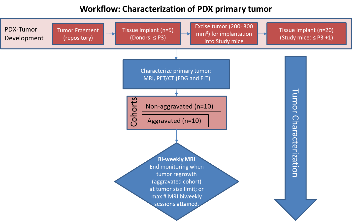
In this study we performed a detailed imaging characterization (workflow below) of this model, details are provided in the attached standard operating procedures. Tumors in half of the mice were resected in the range 200-300 mm3 size; tumors in the other half were allowed to grow until it was necessary to euthanize them because of tumor size.
T2w MRI at 56 days post implant demonstrated a heterogenous tumor with an apparent central hemorrhage and the implant appears to have a defined capsule. On day 98 (post-implant) the tumor increased in size (618%) with very similar imaging characteristics as displayed on day 56.
In contrast, the regrowth following surgical resection of the primary xenograft (105 days post resection) is homogenous, but also appears to have a well-defined capsule.
PET/CT Characterization of the primary tumor: Baseline PET (SOP attached) were performed when tumor reached an approximate 200 mm3. Average SUVbw_max values (n=5) were calculated; [18F]FDG: 1.8 ± 0.2 and [18F]FLT: 2.8 ± 0.7.
The imaging characteristics of this model, which is available from the National Cancer Institute Patient-Derived Models Repository (https://pdmr.cancer.gov/), is highly favorable for preclinical research studies when used in conjunction with non-contrast T2 weighted MRI.
Acknowledgements
We would like to acknowledge the individuals and institutions that have provided data for this collection:
Data Access
| Title | Data Type | Format | Access Points | License | |||
|---|---|---|---|---|---|---|---|
| Images | SR, MR | DICOM | Requires NBIA Data Retriever |
100 | 180 | 2,980 | CC BY 3.0 |
| Standard Operating Procedure 50101: MRI T2 Weighted Non-Contrast Protocol: Single Mouse Pulmonary Gated and Multi-Mouse Non-Gated | CC BY 3.0 | ||||||
| Standard Operating Procedure 50102: Positron Emission Tomography imaging protocol | PET | CC BY 3.0 |
Additional Resources for this Dataset
The National Cancer Institute (NCI) has developed a national repository of Patient-Derived Models (PDMs) comprised of patient-derived xenografts (PDXs), in vitro patient-derived tumor cell cultures (PDCs) and cancer associated fibroblasts (CAFs) as well as patient-derived organoids (PDOrg). These models serve as a resource for public-private partnerships and for academic drug discovery efforts. These PDMs are clinically-annotated with molecular information and made available in the Patient-Derived Model Repository. Data related to the specific subjects in this Collection can be found at:
Click the Versions tab for more info about data releases.
Detailed Description
In addition to images, this collection includes Raw Data Storage SOP Class instances with MR Modality, generated by a Philips MR scanner; this data is not useful to anyone without the proprietary software to interpret it.
Citations & Data Usage Policy
Data Citation |
|
|
Tatum, J., Kalen, J., Ileva, lilia, Riffle, L., Keita, S., Patel, N., Jacobs, P., Sanders, C., James, A., Difilippantonio, S., Thang, L., hollingshead, melinda, Phillips, J., Evrard, Y., Clunie, D., Yanling, Smith, K., Wagner, U., Freymann, J., … Doroshow, J. (2020). Imaging tissue characterization of a patient derived xenograft model of adenocarcinoma pancreas: (PDMR-833975-119-R) [Data set]. The Cancer Imaging Archive. https://doi.org/10.7937/TCIA.0ECK-C338 |
TCIA Citation |
|
|
Clark, K., Vendt, B., Smith, K., Freymann, J., Kirby, J., Koppel, P., Moore, S., Phillips, S., Maffitt, D., Pringle, M., Tarbox, L., & Prior, F. (2013). The Cancer Imaging Archive (TCIA): Maintaining and Operating a Public Information Repository. In Journal of Digital Imaging (Vol. 26, Issue 6, pp. 1045–1057). Springer Science and Business Media LLC. https://doi.org/10.1007/s10278-013-9622-7 |
Other Publications Using This Data
TCIA maintains a list of publications which leverage TCIA data. If you have a manuscript you’d like to add please contact the TCIA Helpdesk.