PDMR-292921-168-R | Imaging characterization of a metastatic patient derived model of adenocarcinoma pancreas:
DOI: 10.7937/TCIA.2020.PCAK-8Z10 | Page Accessibility: Public | Collection
| Location | Species | Subjects | Data Types | Cancer Types | Size | Status | Updated | |
|---|---|---|---|---|---|---|---|---|
| Abdomen | Mouse | 20 | MR, SR | Adenocarcinoma Pancreas | Clinical | Public, Complete | 2023/09/13 |
Summary
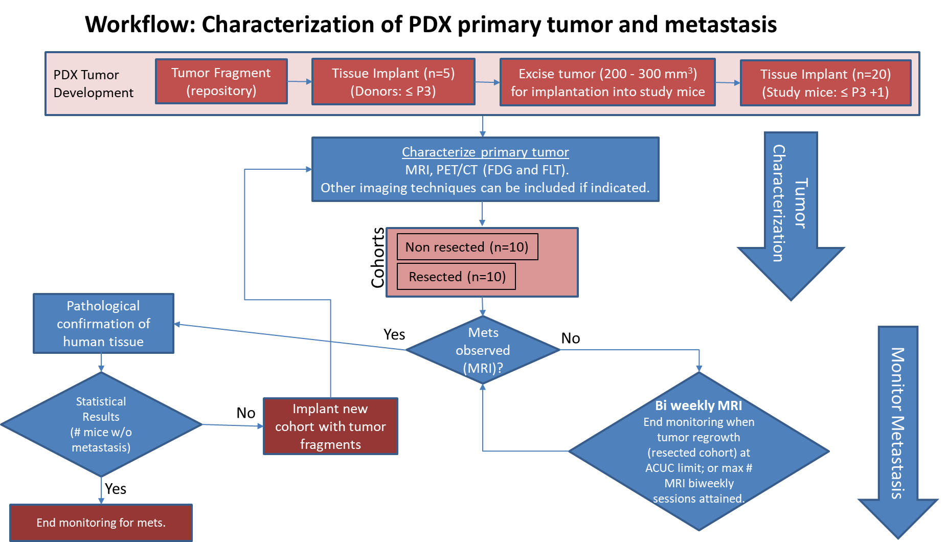
Pre-clinical animal models of spontaneous metastatic cancer are infrequent; the few that exist are resource intensive because determination of the presence of metastatic disease, metastatic burden, and response to therapy normally require multiple timed cohorts with animal sacrifice and extensive pathological examination. We identified and characterized a patient derived xenograft model with metastatic potential, adenocarcinoma pancreas xenograft 292921-168-R. In this study we performed a detailed imaging characterization (workflow below) of this model, which develops spontaneous lung metastases, details are provided in the attached standard operating procedures. Tumors in half of the mice were resected in the range 200-300 mm3 size; tumors in the other half were allowed to grow until it was necessary to euthanize them because of tumor size.
The imaging characteristics of this model (PDMR-292921-168-R), which is available from the National Cancer Institute Patient-Derived Models Repository (https://pdmr.cancer.gov/), is highly favorable for preclinical research studies of metastatic disease when used in conjunction with non-contrast T2 weighted MRI.
Results: Adenocarcinoma pancreas (PDMR-292921-168-R)
Table 1: Penetrance and location of pathological confirmed metastatic lesion(s).
# animals in Group | # animals that displayed metastasis in MRI and confirmed by Pathology | Pathology confirmation of MRI (primary imaging site) | Other confirmed Location (s) | Mouse ID: MRI with pathology confirmation of metastasis |
10 (non-resected) | 4 (5 mice were EU due to xenograft size prior to observation of metastases) | Lung | Lung | 1409, 1410, 1418, 1423 |
10 (resected) | 10 | Lung | Lung, Kidney, Nodes, Peritoneal Wall | 1405, 1407, 1408, 1411, 1413,1415, 1416, 1419, 1420, 1422 |
Percent penetrance with respect to the average time-to-metastasis for non-resected (plot A: time from implant) and resected (plot B: time from tumor resection) cohorts.
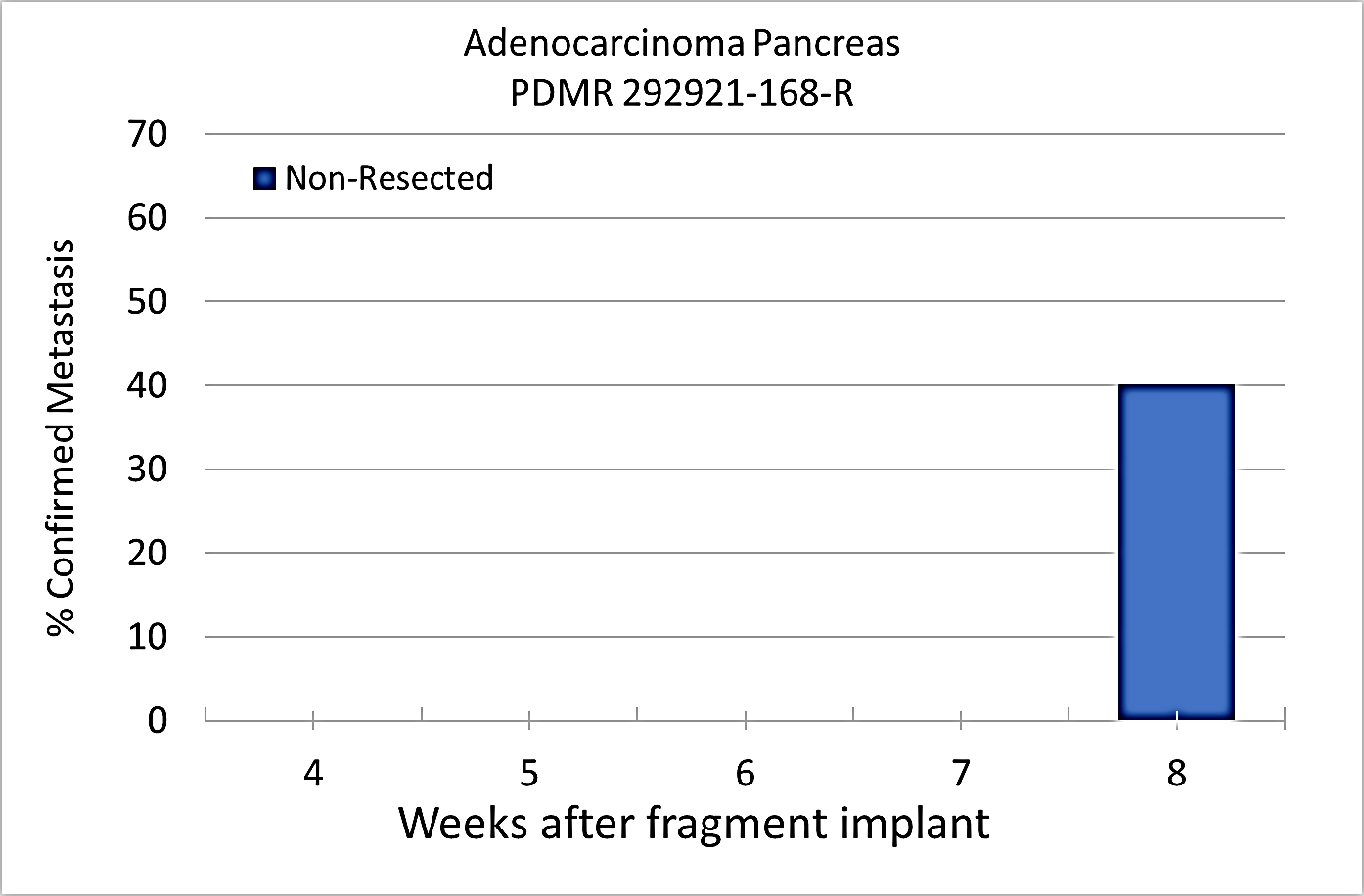 Plot A | 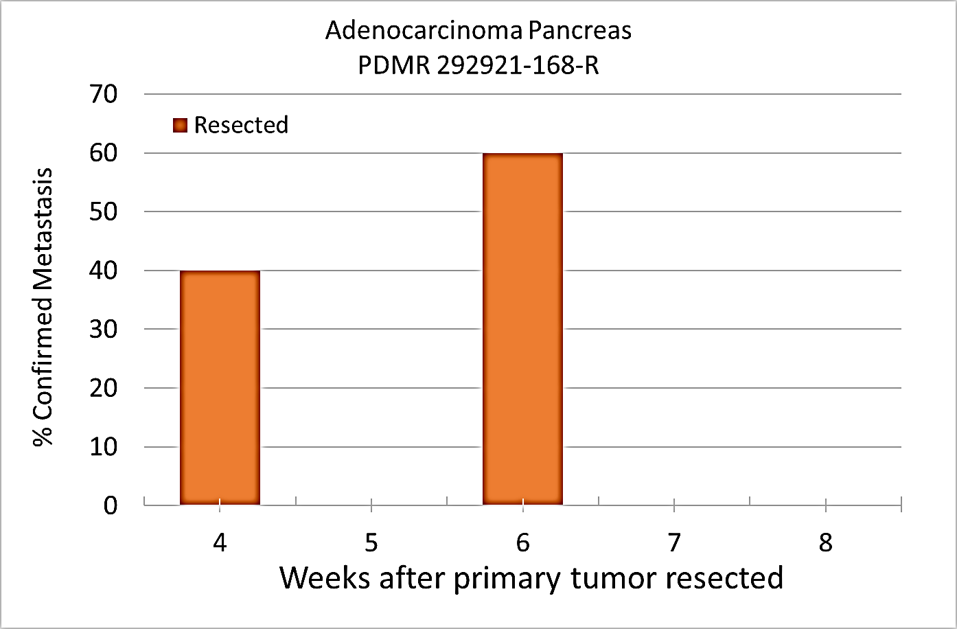 Plot B |
|---|
PET/CT Characterization of the primary tumor: Baseline PET (SOP available below) were performed when tumor reached an approximate 200 mm3. Average SUVmax values (n=5) were calculated; [18F]FDG: 1.3 ± 0.2 and [18F]FLT: 2.3 ± 0.6.
Conclusion
Excellent metastatic model with at least 50% penetrance un-resected and 100% with planned early resection. Metastases are well observed on T2 MRI imaging allowing non-invasive evaluation in treatment trials.Acknowledgements
We would like to acknowledge the individuals and institutions that have provided data for this collection:
Data Access
Click the Versions tab for more info about data releases.
| Title | Data Type | Format | Access Points | License | |||
|---|---|---|---|---|---|---|---|
| Images | SR, MR | DICOM | Requires NBIA Data Retriever |
89 | 160 | 2,657 | CC BY 4.0 |
| Standard Operating Procedure 50101: MRI T2 Weighted Non-Contrast Protocol: Single Mouse Pulmonary Gated and Multi-Mouse Non-Gated | CC BY 4.0 | ||||||
| Standard Operating Procedure 50102: Positron Emission Tomography imaging protocol | PET | CC BY 4.0 |
Additional Resources for this Dataset
The National Cancer Institute (NCI) has developed a national repository of Patient-Derived Models (PDMs) comprised of patient-derived xenografts (PDXs), in vitro patient-derived tumor cell cultures (PDCs) and cancer associated fibroblasts (CAFs) as well as patient-derived organoids (PDOrg). These models serve as a resource for public-private partnerships and for academic drug discovery efforts. These PDMs are clinically-annotated with molecular information and made available in the Patient-Derived Model Repository. Data related to the specific subjects in this Collection can be found at:
The NCI Cancer Research Data Commons (CRDC) provides access to additional data and a cloud-based data science infrastructure that connects data sets with analytics tools to allow users to share, integrate, analyze, and visualize cancer research data.
- Imaging Data Commons (IDC) (Imaging Data)
Detailed Description
In addition to images, this collection includes Raw Data Storage SOP Class instances with MR Modality, generated by a Philips MR scanner; this data is not useful to anyone without the proprietary software to interpret it.
Citations & Data Usage Policy
Data Citation |
|
|
Tatum, J., Kalen, J., Ileva, lilia, L, R., S, K., N, P., Jacobs, P., Sanders, C., A, J., Difilippantonio, S., L, T., hollingshead, melinda, J, P., Y, E., Clunie, D., Y, L., Suloway, C., Smith, K., U, W., … Doroshow, J. (n.d.). Imaging characterization of a metastatic patient derived model of adenocarcinoma pancreas: (PDMR-292921-168-R). The Cancer Imaging Archive. https://doi.org/10.7937/TCIA.2020.PCAK-8Z10 |
TCIA Citation |
|
|
Clark, K., Vendt, B., Smith, K., Freymann, J., Kirby, J., Koppel, P., Moore, S., Phillips, S., Maffitt, D., Pringle, M., Tarbox, L., & Prior, F. (2013). The Cancer Imaging Archive (TCIA): Maintaining and Operating a Public Information Repository. In Journal of Digital Imaging (Vol. 26, Issue 6, pp. 1045–1057). Springer Science and Business Media LLC. https://doi.org/10.1007/s10278-013-9622-7 |
Other Publications Using This Data
TCIA maintains a list of publications which leverage TCIA data. If you have a manuscript you’d like to add please contact the TCIA Helpdesk.